
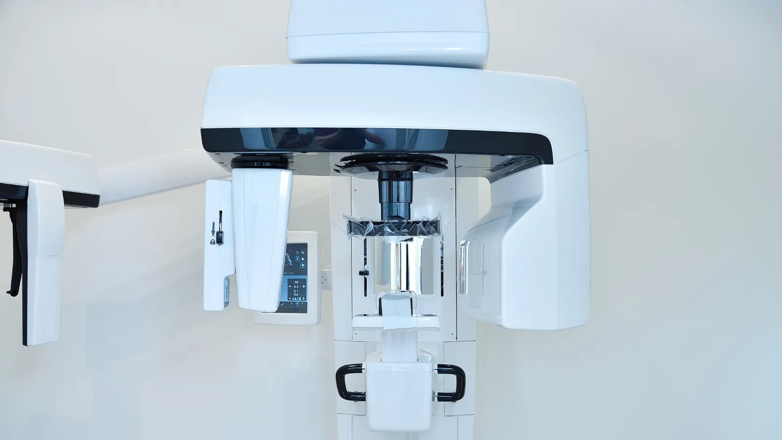
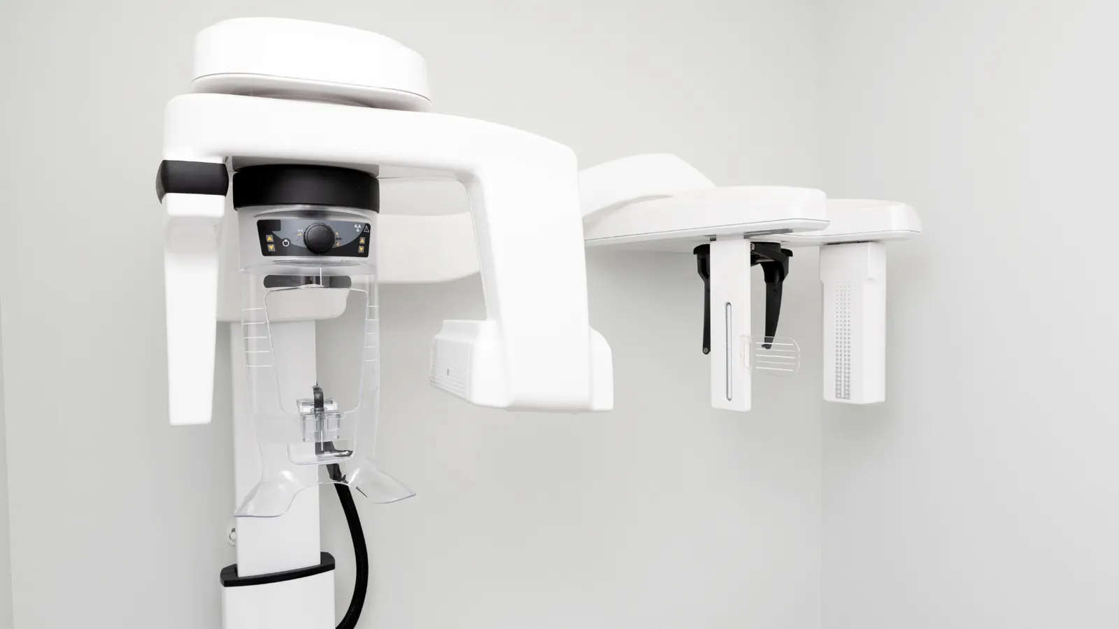
A dental CT scan produces a three dimensional image of the teeth, the jawbone and the structures around them. A conventional dental X-ray flattens everything into a single plane; a CT scan keeps the depth, so a site can be looked at from any direction and measured. At Dentopia the scanner is on site, which means the images are taken and read as part of planning rather than ordered elsewhere and waited for.
What the images show
The scan shows the shape and volume of the jawbone and how dense it is, which is what determines whether a site can hold an implant. It shows the position of the structures that surgery has to respect, among them the sinus cavities and the nerve canals. It also brings out findings that a flat image can hide, such as cysts in the jaw, and it shows the full shape of a root, including canals that curve or divide.
When a scan is used
Implant planning is the most common reason: the position, the angle and the depth of an implant are decided on the scan before anything is placed. It is used before removing a wisdom tooth that sits close to a nerve canal, so that the approach is known in advance. In root canal treatment it helps where a tooth has not settled and the anatomy needs to be seen properly. A scan is requested when it changes a decision, not added to every examination as a matter of course.
How the examination goes
You sit or stand with your chin on a support while the scanner turns once around your head. Nothing goes into the mouth and nothing touches the teeth. It takes a matter of seconds, you are asked only to hold still, and the images are ready straight afterward for the dentist to go through with you.



Book this treatment
Pick a time that suits you. We will call you back to confirm the appointment.